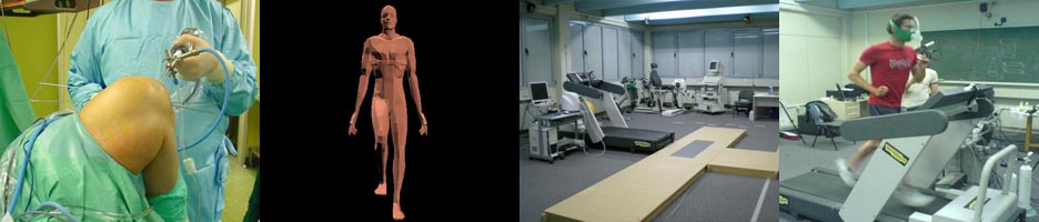
Ορθοπαιδικό Αθλητιατρικό Κέντρο Ιωαννίνων

“Ένα σύγχρονο και πλήρως εξοπλισμένο εργαστήριο”

The Diagnostic Dilemma caused by osteoid osteoma that presents as knee pain.

Georgoulis A., Papageorgiou C., Moebius U., Rossis J., Papadonikolakis A., Soucacos P.N.

Arthroscopy, 18(1):32-37, 2002

PURPOSE: Osteoid osteoma is a rare benign bone lesion with a high incidence in adolescents and young people. The objective of our study was to illustrate the difficulties in diagnosis of osteoid osteoma in patients presenting with atypical knee pain.

TYPE OF STUDY: Retrospective case series.

METHODS: In 10 patients who presented to our department with atypical knee pain between 1984 and 1999, the diagnosis of an osteoid osteoma was delayed. Retrospective review of these 10 cases was performed using interviews and re-evaluation of medical histories, radiographs, computed tomography (CT) scans, isotope bone scan, and magnetic resonance imaging (MRI).

RESULTS: Initial radiographs showed features of osteiod osteoma in only 2 cases. In addition, in 1 case, not only initial but also repeated radiographs of the knee joint were still normal 10 months after the delineation of the nidus using MRI. Four unnecessary arthroscopies were performed on 4 of the 10 patients and the final diagnosis was established using MRI, CT, and isotope bone scan. The mean time interval between arthroscopy and osteiod osteoma diagnosis was 11.5 months.

CONCLUSIONS: Osteoid osteoma must be included in the differential diagnosis of persistent unexplained knee pain, especially when objective findings of the knee are vague. The presence of the lesion juxta-articular to the knee joint or in the midshaft or upper end of the femur may be referred as pain to a nearby joint. Plain radiographs have a low diagnostic value in the detection of the lesion whereas isotope bone scan and MRI are reliable imaging techniques. The evaluation of the ipsilateral hip joint should not be overlooked.